Talar Neck Fracture Specialists
Do you participate in high-impact sports or activities? If so, you may be at an increased risk of experiencing a talar neck fracture. The talus is the primary ankle bone and is irregularly shaped, with a head, body, and neck. Fractures to the neck portion of the brain can cause pain, swelling, and an inability to put weight on the foot. Foot and ankle specialists, Doctors Thomas Haytmanek and Jonathon Backus have diagnosed many patients in Vail and Frisco, Colorado, as well as Denver, Boulder, and surrounding Summit County areas who have experienced a talar neck fracture. For a specialized treatment plan, contact The Steadman Clinic’s Sports Foot and Ankle team today!

What is a talar neck fracture?
The talus is a small bone in the foot that sits between the tibia (shin bone) and the calcaneus (heel bone). It is thought of as the “ankle bone”. The talus has an irregular dome shape like a turtle shell. The talus is broken up into three sections: head, neck and body. A talar neck fracture describes the location where the ankle bone is broken. A talar neck fracture occurs when a crack or break appears in the juncture between the head and the neck. A talar neck fracture happens when a major force drives the foot into the ankle from high energy accidents like car accidents or skiing and snowboarding. Our physicians, orthopedic foot and ankle specialists, treat patients from Vail, Colorado, and the surrounding Eagle and Summit Counties who have suffered a talar neck fracture. The talus is a tricky bone, as it is mostly covered by cartilage and the blood supply is unusually tenuous. We have extensive experience in successfully treating foot and ankle injuries and helping to return patients to their pre-injury lifestyle.
CLICK IMAGE TO ENLARGE
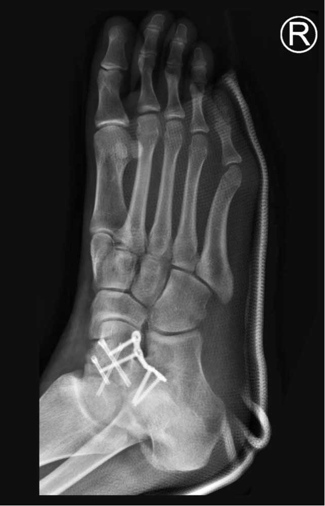
Talar Neck Fracture
What are the symptoms of a talar neck fracture?
Symptoms of the broken ankle or broken talus in particular include:
- Pain throughout the foot and ankle
- Inability to bear weight
- Swelling and tenderness
It is possible for the broken bone to puncture through the skin when the injury happens. The foot may appear twisted or deformed. It is likely the bone will be displaced, requiring surgery.
How is a talar neck fracture diagnosed?
Our physicians will perform a physical examination of the injured area and will order x-rays. If the fracture is not displaced, it can be difficult to see on an x-ray. Our physicians may order a CT scan or an MRI. The CT scan can help him visualize and access the fracture more easily and determine if surgery is required. An MRI can show surrounding soft tissue injury well.
What is the treatment for a talar neck fracture?
Non-Surgical Treatment:
Although rare for a talar neck fracture to not be displaced, it is possible to heal the break by immobilizing the injury with a cast below the knee and refrain from bearing weight on it for a six to eight-week period. Once imaging confirms the fracture is healed, the patient will be allowed to bear weight. Prescribed exercises can help improve strength and mobility after the bone is headed.
Surgical Treatment:
Surgery is also called open reduction- internal fixation (ORIF). Surgery is always necessary if the bone is displaced and if there is concern the displacement may be disrupting the blood supply. During surgery, the talus will be repositioned to its original placement within the ankle. Different approaches can be used and a combination of screws or plates and screws can be used to stabilize the fracture. Occasionally, dissolving pins are used to stabilize the fracture. After surgery, the patient is typically splinted (a soft cast) for the first two weeks. Patients remain non-weight bearing for approximately six to eight weeks. Early range of motion may be allowed depending on the severity of the injury and the fixation used.
For more information on talar neck fractures and the treatment options available, please contact the office of Sports Foot and Ankle serving Vail Colorado and the surrounding Eagle and Summit counties.

