Calcaneofibular Ligament Tear Specialists
Are you an athlete who participates in skiing, snowboarding, or other similar sports? If so, you may be at risk of sustaining a high-impact injury from a hard twisting fall. Injuries of this type can cause the calcaneofibular ligament to tear. A tear to this ligament, whether partially or completely, has a significant impact on its ability to provide ankle joint stability. Dr. Thomas Haytmanek and Dr. Jonathan Backus specialize in treating calcaneofibular ligament tears for patients from Vail and Frisco, Colorado, as well as Denver, Boulder, and surrounding Summit County areas. Contact The Steadman Clinic’s Sports Foot and Ankle team today!

What is a calcaneofibular ligament tear?
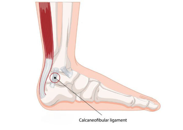
The calcaneofibular ligament (CFL) is one of three lateral ankle ligaments that connects the lateral malleolus of the fibula to the lateral surface of the calcaneus. This ligament’s primary role is to prevent exaggerated inversion of the subtalar joint (a common type of ankle injury). High-impact injuries from a twisting injury, fall, or ski/snowboard crash can cause the calcaneofibular ligament to tear. A tear to this ligament, whether partially or completely, has a significant impact on its ability to provide ankle joint stability. Dr. Thomas Haytmanek and Dr. Jonathan Backus specialize in treating calcaneofibular ligament tears for patients from Vail and Frisco, Colorado, as well as Denver, Boulder, and surrounding Summit County areas.

What are the symptoms of a calcaneofibular ligament tear?
Individuals with a calcaneofibular ligament tear most commonly report pain and swelling along the outer ankle. These symptoms can vary depending on the severity of the traumatic event. Individuals with partial calcaneofibular ligament tears may not experience as significant of symptoms as those with completely ruptured calcaneofibular ligaments. Some other common symptoms of a calcaneofibular ligament tear include:
- Ankle instability, or a feeling that the ankle “gives out”
- Worsening pain with weight-bearing activities
- Bruising at the site of injury
How is a calcaneofibular ligament tear diagnosed?
The patient’s medical history is gathered followed by a thorough physical examination. The integrity of each ankle ligament is evaluated through a variety of physical exam maneuvers. A positive anterior drawer and/or talar tilt test is suggestive of a lateral ligament tear, such as the calcaneofibular ligament. Many patients also sprain the calcaneofibular ligament and posterior talofibular ligament at the same time. Magnetic resonance imaging (MRI) is also utilized to confirm a calcaneofibular ligament tear. These advanced imaging studies can also identify the degree of damage to these soft tissue structures.
What is the treatment for a calcaneofibular ligament tear?
Non-surgical treatment:
Patients with a calcaneofibular ligament tear without evidence of an ankle fracture can be treated with conservative therapies alone. It is important for the ankle joint motion to be controlled to allow for ligament healing. A tall walking boot is typically used for this immobilization. Physical therapy is also utilized to strengthen the ankle joint. The patient’s specific injury will determine the length of the immobilization period as well as the number of physical therapy sessions. Any pain and inflammation can be reduced through rest, ice, elevation, and non-steroidal anti-inflammatory medications (NSAIDs).
Surgical treatment:
Surgical intervention is often necessary for patients with continued ankle joint instability after conservative management or some patients who experienced a complete rupture of the calcaneofibular ligament. Surgical intervention may be needed to repair the calcaneofibular ligament tear. The damaged portions of the ligament are cleaned and the remaining healthy tissue is sutured back together or reattached to the bone. The exact incision and technique used depend on the nature of the tear. A tendon graft may be required for ligament reconstruction in patients with irreparable damage to the calcaneofibular ligament.
